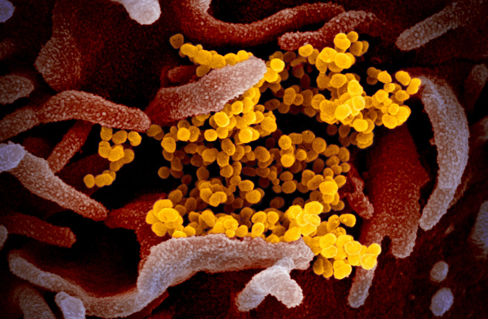
We now have microscopic shots of the novel coronavirus (SARS-CoV-2, previously referred to as 2019-nCoV) that has been spreading around China and the world.
The National Institute of Allergy and Infectious Diseases (NIAID) in the US released new images of the novel coronavirus on Thursday, February 13. The institute’s Rocky Mountain Laboratories based in Hamilton, Montana produced the images on its scanning and transmission electron microscopes, according to a post on NIAID’s official website.
The virus, which causes the disease known as Covid-19, was first detected in Wuhan in December 2019, and has developed into a global public health emergency. As of press time, over 60,000 total cases have been confirmed, with 1,381 patients dead and 6,809 cured.
Rocky Mountain Laboratory investigator Emmie de Wit, Ph.D., provided the virus samples to the lab as part of her research and microscopist Elizabeth Fischer was able to produce the images. The images were digitally colorized by the lab’s visual medical arts office – giving the virus a pretty neat aesthetic, we’d like to add.
The images are said to be comparable to SARS-CoV (severe acute respiratory syndrome coronavirus) and MERS-CoV (Middle East respiratory syndrome coronavirus) which emerged in 2002 and 2012, respectively. In their post, NIAID said that the similarities are “not surprising: The spikes on the surface of coronaviruses give this virus family its name – corona, which is Latin for “crown,” and most any coronavirus will have a crown-like appearance.”
Check out images of the virus below:
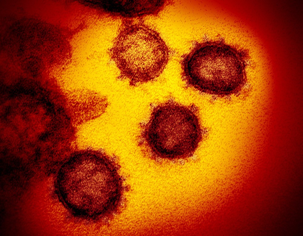 Image via NIAID-RML
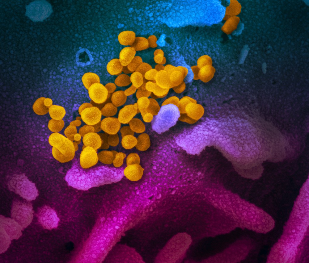
Image via NIAID-RML
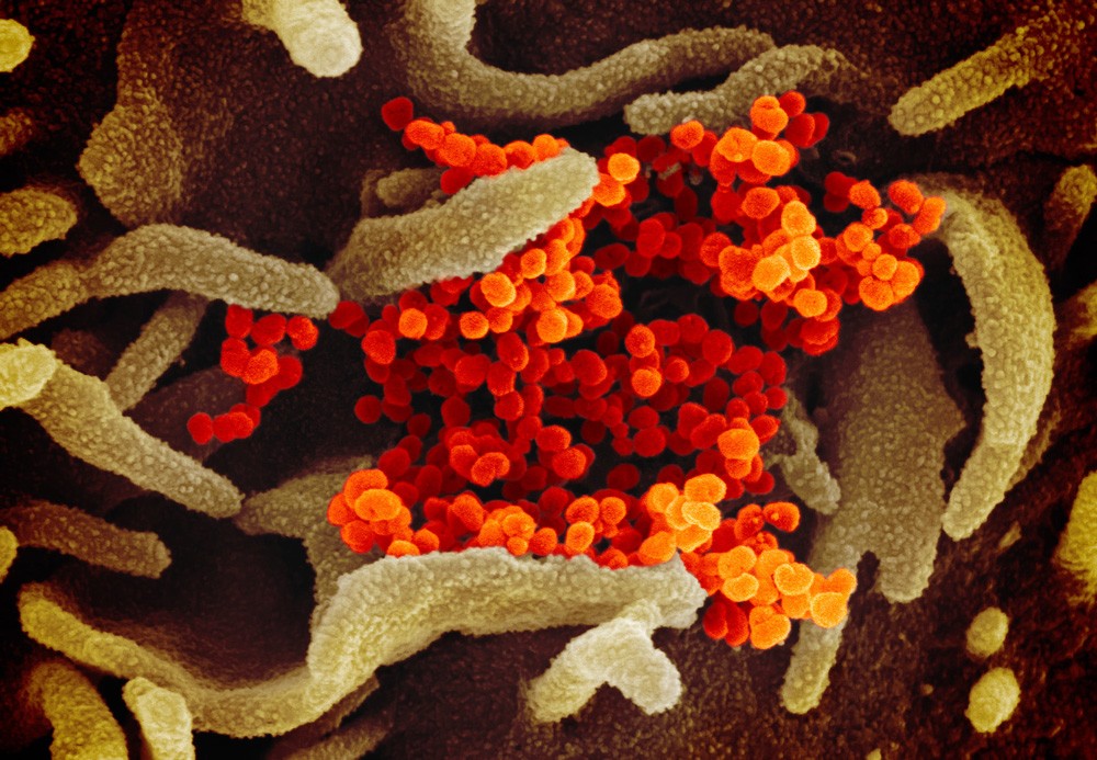
Image via NIAID-RML
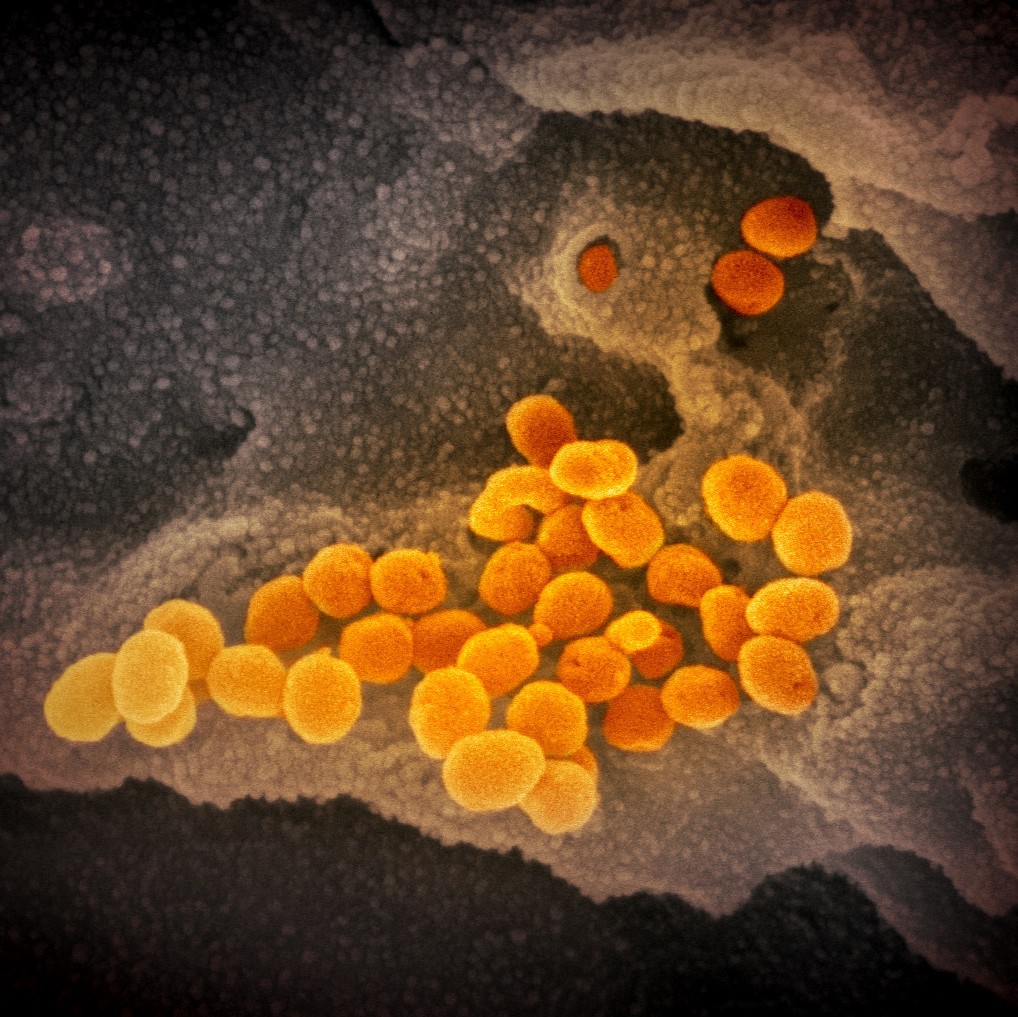
Image via NIAID-RML
For regular updates on the novel coronavirus outbreak in China, .
[Cover image via NIAID-RML]



Recent Comments